BSOS and the Brain: Research and Impact

As social scientists, BSOS researchers don’t just study how people think and act, and the systems within which they do so. They also explore what drives human behavior, sometimes down to a neurological level.
Read on to learn about our community’s recent contributions to brain research.
Tracy Riggins 
PSYC Professor
Riggins spent the 2024-2025 academic year focusing on two significant brain-related projects during sabbatical leave. She wrote a paper on current knowledge on the neural bases of memory development (the last paper on the topic was published more than 10 years ago). She also created a neuroanatomical template that researchers can use to segment parts of the hippocampus from infant MRIs.
Both efforts were aided by Riggins’ past, and present, research studies on child development, memory and cognition. Two are still enrolling participants. One is designed to track the brain development, memory performance and nap status of 180 children ages 3 to 4 1/2 over the course of one year. This study (go.umd.edu/NapStudy) focuses on what cognitive, physiological and neural changes accompany children transitioning from taking a midday nap to no nap at all.
The second project is the HEALthy Brain and Child Development (HBCD) Study (hbcdstudy.org), which tracks individuals in their second trimester of pregnancy and follows them and their children through early childhood to establish a baseline for typical brain development. This research provides insights on factors that could affect brain development, such as positive relationships with caregivers, poverty, social and emotional support, and more.
Paul LaFosse

Graduate Student, Program in Neuroscience and Cognitive Science (NACS)
LaFosse’s recently published, coauthored research set out to measure the input-output function of neurons—the transformation neurons go through when they receive a signal and generate a response, known as the neurons’ spike rate—in conscious mice. Past studies have only measured this in mice in anesthetized states, or cells in vitro.
Using light to control brain cell activity in an awake mouse—by directing light at many neurons at the same time to make them fire—the researchers were able to see that input-output functions can change as brain state changes, like being awake or being asleep. They also found that neurons rest right at the transition point of the function’s shape, between a straight region and a curved (nonlinear) region.
“This means that when inputs arrive to the brain that make a cell less active, this decreases the effect of other inputs even more, resulting in a feedback loop that can dramatically suppress or filter those inputs,” said LaFosse.
LaFosse and his collaborators said this study helps to shed light not only on brain function, but the input/output processes in Artificial Intelligence and Machine Learning uses, including ChatGPT.
Weizhen Xie

PSYC Assistant Professor
Xie recently concluded a study that is among the first to show that when humans have to identify and categorize an image, like seeing a dog and recognizing it as an animal rather than a human, it’s not just about how many times brain neurons fire. The order in which individual neurons fire also plays an important part in the process.
To do this, Xie and colleagues had eight treatment-resistant epilepsy patients—who had had electrodes implanted into their brain to identify where their seizures were happening—participate in a task that involved them putting an image into one of four categories: animal, object, person or place.
The researchers found that the sequence of neuronal activity during the task offers new insights into brain function, and has implications for advancing artificial intelligence.
“Understanding how the human brain categorizes things so much more efficiently could help develop AI models that learn faster and use far less energy,” said Xie.
Evan Hart

PSYC Assistant Professor
Since joining UMD in 2024, Hart has been working to finalize and publish the results of his postdoctoral research project on how the brain takes in sensory information to help someone decide between going one way to work or another, and which duties to perform once they arrive, for example.
To study this, Hart implanted six rats with microelectrodes and trained them to make different responses to 96 distinct odors that they were presented with. Each odor informed rats to perform one of two responses, rewarded with tropical punch- or grape-flavored water.
Hart recorded the rats’ brain neurons’ spiking patterns and found that the part responsible for this sort of decision-making is the orbitofrontal cortex, where neural activity is highly flexible—so much so that it is able to rank sensory information, like smells, according to importance.
This research could help us to better understand how the brain makes complex decisions while thinking about the future, and—given the predictable nature of the trained rats’ responses to certain smells—even the best way for the human brain to learn.
Caroline J Charpentier

PSYC Assistant Professor
Charpentier recently launched the second phase of a National Institute of Mental Health-funded research study to better understand various factors that contribute to social learning, the process through which individuals gain knowledge from other people.
Charpentier and her team have already gained initial insights into social learning behavior through an online study in which 800 participants from the general population completed cognitive tasks measuring how they learned through observation, how trust impacted their learning, and how risky decisions were influenced by peers. They also asked participants questions about mental health symptoms and metrics related to autism, to understand how social learning varies with social difficulties experienced in daily life.
The next phase of the project will examine what happens in the brain when neurodiverse individuals (including those with and without autism) complete the same social learning tasks in an MRI scanner, providing insights into how brain activity during social learning relates to neurodiversity. The project should conclude in March 2027.
Xuan (Anna) Li

PSYC Assistant Professor
With Najib El-Sayed, a professor of cell biology and molecular genetics and the University of Maryland’s Institute for Advanced Computer Studies, Li is working on a UMD Brain and Behavior Institute-funded study of how gene expression and epigenetic factors—which turn genes on and off without changing genetic information itself—play a part in opioid relapse.
Using rats, Li is using non-harmful viral vectors to tag the brain circuit of interest and training rats to take opioids voluntarily. Li and El-Sayed will then examine changes in gene expression, and how genes turn on and off after a period of abstinence from opioid use.
Results of the study, expected later this summer, will help us to understand what molecular changes are associated with persistent relapsing on opioids—and, perhaps, identify potential therapeutic targets to promote abstinence and prevent relapse.
Elizabeth Redcay

PSYC Professor
Redcay and colleagues are working to understand how the brain supports social interactions. The goal of this Brain and Behavior Institute-funded study—which Redcay is working on with PSYC Assistant Professor Caroline Charpentier, Assistant Professor Rachel Romeo, who has a joint appointment in HESP and the Department of Human Development and Quantitative Methodology and Philip Resnik, a professor in the Department of Linguistics and the University of Maryland Institute for Advanced Computer Studies—is to see how people’s brain activity and behaviors (e.g., phrases, facial expressions, and body gestures) align with each other or “get in sync” during natural social interactions.
To do this, 30 pairs of study participants will wear caps, called near-infrared spectroscopy (fNIRS) devices, that measure changes in the brain while the pairs interact with one another in real-time. Partners will do interactive cooperative, competitive, and communication games like rock-paper-scissors, as well as independent tasks that measure brain activity when thinking about social goals. Then, to validate fNIRS results, roughly 20 study participants will undergo a functional magnetic resonance imaging (fMRI), which provides much greater detail on which brain areas are active while doing some of the same tasks from the fNIRS portion.
Findings from the study will help shed new light on the biological drivers of human communication and connection.
Alexander Shackman & Jack Blanchard

PSYC Professors
“The treatments we have at present for people with psychosis spectrum disorders are far from curative,” said Shackman. “Current treatments mainly help with cognitive deficits, to think clearly or stay on track, but they don’t help with some of the major emotional symptoms that characterize people living with these conditions.”

A study that Shackman and Blanchard plan to wrap up later this year seeks to help change that. Using fitbit-like actigraphs to measure participants’ sleep quality, and smartphones to monitor their geolocation, symptoms, and mood multiple times per day for one week, the researchers are able to assess participants’ in-the-moment paranoia. They are also able to see what’s going on in the brain when these feelings occur, as participants are then asked to come in for an fMRI scan that asks them to complete multiple different tasks, including watching a movie and being virtually led down a hallway that ends either with something scary hiding behind the door, or nothing at all, 50% of the time.
The researchers hope that by seeing what factors and scenarios prompt some of the emotional symptoms of paranoia, and in what parts of the brain those symptoms originate, findings from the study will one day lead to better treatments for people with psychosis spectrum disorders.
Adam Brockett, PSYC ’11
Former Assistant Research Professor
Before he began working as an assistant professor at the University of New Hampshire in 2024, Brockett completed a study at UMD showing how the brain is impacted by a psychedelic drug being studied to potentially treat depression: psilocybin, a component of hallucinogenic “magic mushrooms.”
Through a UMD Grand Challenges Grant Program award, Brockett examined the behavior and auditory cortex of live mice before and after a dose of psilocybin. He found that, about 10 minutes after the mice were given psilocybin, their neurons became very quiet. Thirty minutes after receiving the drug, the functional connectivity between the mice’s brain regions—how the parts of the brain interact with each other—increased.
“What might be happening when psilocybin is taken is that subjects become less aware of what's around them, and more focused on some sort of internal change or sensation that they might be experiencing,” said Brockett. “This gives us a baseline understanding of what these drugs are actually doing to our senses. I think this helps us kind of map that space, and I think the more that we have this mapped out, the more likely that it becomes a tenable option for people.”
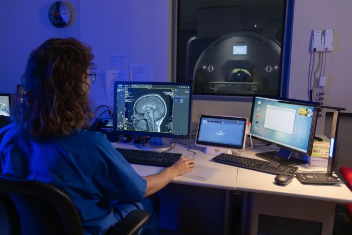
Photo of the Maryland Neuroimaging Center taken by Tom Bacho
Published on Wed, Apr 2, 2025 - 10:40AM


